 PDF(1256 KB)

Research progress on the active ingredients of traditional Chinese medicine against hepatic fibrosis by regulating the MAPK signaling pathway
HUANG Fujing, XIE Xingxing, LIU Xue, FAN Ling, YU Zhigang, ZHANG Jinjuan
Chinese Journal of Hospital Pharmacy ›› 2025, Vol. 45 ›› Issue (2) : 225-233.
PDF(1256 KB)

 PDF(1256 KB)
Research progress on the active ingredients of traditional Chinese medicine against hepatic fibrosis by regulating the MAPK signaling pathway
 ({{custom_author.role_en}}), {{javascript:window.custom_author_en_index++;}}
Hepatic fibrosis (HF) is a reversible pathological process associated with various chronic liver diseases. If left untreated, it can progress to severe pathological stages like cirrhosis and liver cancer, significantly impacting the health and quality of life. Currently, available clinical treatments for HF demonstrate limited efficacy, highlighting the urgent need for new drugs or active ingredients against HF. The mitogen-activated protein kinase (MAPK) signaling pathway plays a crucial regulatory role in cell proliferation, differentiation, and apoptosis, making it a key target in the modulation of HF development. Research has shown that interfering with the activation of MAPK signaling pathways through active ingredients of traditional Chinese medicine (TCM) herbals can yield positive outcomes in combating HF. This review examined the role of the MAPK signaling pathway in regulating HF and explored the therapeutic potential of active ingredients of TCM herbals targeting this pathway. Our aim is to provide insights that may contribute to the future development of novel drugs against HF.
hepatic fibrosis / MAPK signaling pathway / active ingredients of traditional Chinese medicine / research progress {{custom_keyword}} /
| 1 |
{{custom_citation.content}}
{{custom_citation.annotation}}
|
| 2 |
{{custom_citation.content}}
{{custom_citation.annotation}}
|
| 3 |
Liver fibrosis is a major cause of morbidity and mortality worldwide due to chronic viral hepatitis and, more recently, from fatty liver disease associated with obesity. Hepatic stellate cell activation represents a critical event in fibrosis because these cells become the primary source of extracellular matrix in liver upon injury. Use of cell-culture and animal models has expanded our understanding of the mechanisms underlying stellate cell activation and has shed new light on genetic regulation, the contribution of immune signaling, and the potential reversibility of the disease. As pathways of fibrogenesis are increasingly clarified, the key challenge will be translating new advances into the development of antifibrotic therapies for patients with chronic liver disease.
{{custom_citation.content}}
{{custom_citation.annotation}}
|
| 4 |
Chronic liver injury leads to liver inflammation and fibrosis, through which activated myofibroblasts in the liver secrete extracellular matrix proteins that generate the fibrous scar. The primary source of these myofibroblasts are the resident hepatic stellate cells. Clinical and experimental liver fibrosis regresses when the causative agent is removed, which is associated with the elimination of these activated myofibroblasts and resorption of the fibrous scar. Understanding the mechanisms of liver fibrosis regression could identify new therapeutic targets to treat liver fibrosis. This Review summarizes studies of the molecular mechanisms underlying the reversibility of liver fibrosis, including apoptosis and the inactivation of hepatic stellate cells, the crosstalk between the liver and the systems that orchestrate the recruitment of bone marrow-derived macrophages (and other inflammatory cells) driving fibrosis resolution, and the interactions between various cell types that lead to the intracellular signalling that induces fibrosis or its regression. We also discuss strategies to target hepatic myofibroblasts (for example, via apoptosis or inactivation) and the myeloid cells that degrade the matrix (for example, via their recruitment to fibrotic liver) to facilitate fibrosis resolution and liver regeneration.
{{custom_citation.content}}
{{custom_citation.annotation}}
|
| 5 |
Multicellular organisms use mitogens to regulate cell proliferation, but how fluctuating mitogenic signals are converted into proliferation-quiescence decisions is poorly understood. In this work, we combined live-cell imaging with temporally controlled perturbations to determine the time scale and mechanisms underlying this system in human cells. Contrary to the textbook model that cells sense mitogen availability only in the G cell cycle phase, we find that mitogenic signaling is temporally integrated throughout the entire mother cell cycle and that even a 1-hour lapse in mitogen signaling can influence cell proliferation more than 12 hours later. Protein translation rates serve as the integrator that proportionally converts mitogen history into corresponding levels of cyclin D in the G phase of the mother cell, which controls the proliferation-quiescence decision in daughter cells and thereby couples protein production with cell proliferation.Copyright © 2020 The Authors, some rights reserved; exclusive licensee American Association for the Advancement of Science. No claim to original U.S. Government Works.
{{custom_citation.content}}
{{custom_citation.annotation}}
|
| 6 |
{{custom_citation.content}}
{{custom_citation.annotation}}
|
| 7 |
周鑫, 王智, 何雪茹, 等. 潜在抗肝纤维化药物与靶点相关信号通路研究进展[J]. 临床肝胆病杂志, 2023, 39(12): 2932-2941.
{{custom_citation.content}}
{{custom_citation.annotation}}
|
| 8 |
{{custom_citation.content}}
{{custom_citation.annotation}}
|
| 9 |
{{custom_citation.content}}
{{custom_citation.annotation}}
|
| 10 |
{{custom_citation.content}}
{{custom_citation.annotation}}
|
| 11 |
{{custom_citation.content}}
{{custom_citation.annotation}}
|
| 12 |
{{custom_citation.content}}
{{custom_citation.annotation}}
|
| 13 |
The Ras-dependent extracellular signal-regulated kinase (ERK)1/2 mitogen-activated protein (MAP) kinase pathway plays a central role in cell proliferation control. In normal cells, sustained activation of ERK1/ERK2 is necessary for G1- to S-phase progression and is associated with induction of positive regulators of the cell cycle and inactivation of antiproliferative genes. In cells expressing activated Ras or Raf mutants, hyperactivation of the ERK1/2 pathway elicits cell cycle arrest by inducing the accumulation of cyclin-dependent kinase inhibitors. In this review, we discuss the mechanisms by which activated ERK1/ERK2 regulate growth and cell cycle progression of mammalian somatic cells. We also highlight the findings obtained from gene disruption studies.
{{custom_citation.content}}
{{custom_citation.annotation}}
|
| 14 |
The proapoptotic protein Bim is expressed de novo following withdrawal of serum survival factors. Here, we show that Bim-/- fibroblasts and epithelial cells exhibit reduced cell death following serum withdrawal in comparison with their wild-type counterparts. In viable cells, Bax associates with Bcl-2, Bcl-x(L) and Mcl-1. Upon serum withdrawal, newly expressed Bim(EL) associates with Bcl-x(L) and Mcl-1, coinciding with the dissociation of Bax from these proteins. Survival factors can prevent association of Bim with pro-survival proteins by preventing Bim expression. However, we now show that even preformed Bim(EL)/Mcl-1 and Bim(EL)/Bcl-x(L) complexes can be rapidly dissociated following activation of ERK1/2 by survival factors. The dissociation of Bim from Mcl-1 is specific for Bim(EL) and requires ERK1/2-dependent phosphorylation of Bim(EL) at Ser(65). Finally, ERK1/2-dependent dissociation of Bim(EL) from Mcl-1 and Bcl-x(L) may play a role in regulating Bim(EL) degradation, since mutations in the Bim(EL) BH3 domain that disrupt binding to Mcl-1 cause increased turnover of Bim(EL). These results provide new insights into the role of Bim in cell death and its regulation by the ERK1/2 survival pathway.
{{custom_citation.content}}
{{custom_citation.annotation}}
|
| 15 |
Senescence is a result of cellular stress and is a potential mechanism for regulating cancer. As a member of the mitogen-activated protein kinase family, ERK1/2 (extracellular signal-regulated protein kinase) has an important role in delivering extracellular signals to the nucleus, and these signals regulate the cell cycle, cell proliferation and cell development. Previous studies demonstrated that ERK1/2 is closely associated with cell aging; however other previous studies suggested that ERK1/2 exerts an opposite effect on aging models and target proteins, even within the same cell model. Recent studies demonstrated that the effect of ERK1/2 on aging is likely associated with its target proteins and regulators, negative feedback loops, phosphorylated ERK1/2 factors and ERK1/2 translocation from the cytoplasm to the nucleus. The present review aims to examine the mechanism of ERK1/2 and discuss its role in cellular outcomes and novel drug development.
{{custom_citation.content}}
{{custom_citation.annotation}}
|
| 16 |
The p38 MAPK (mitogen-activated protein kinase) signalling pathway allows cells to interpret a wide range of external signals and respond appropriately by generating a plethora of different biological effects. The diversity and specificity in cellular outcomes is achieved with an apparently simple linear architecture of the pathway, consisting of a core of three protein kinases acting sequentially. In the present review, we dissect the molecular mechanisms underlying p38 MAPK functions, with special emphasis on the activation and regulation of the core kinases, the interplay with other signalling pathways and the nature of p38 MAPK substrates as a source of functional diversity. Finally, we discuss how genetic mouse models are facilitating the identification of physiological functions for p38 MAPKs, which may impinge on their eventual use as therapeutic targets.
{{custom_citation.content}}
{{custom_citation.annotation}}
|
| 17 |
{{custom_citation.content}}
{{custom_citation.annotation}}
|
| 18 |
{{custom_citation.content}}
{{custom_citation.annotation}}
|
| 19 |
郝少东, 刘彩萍, 李月廷, 等. 肝星状细胞活化相关信号通路在肝纤维化中的研究进展[J]. 胃肠病学和肝病学杂志, 2022, 31(2): 131-135.
{{custom_citation.content}}
{{custom_citation.annotation}}
|
| 20 |
Upon liver injury, hepatic stellate cells (HSC) show increased proliferation, motility, and extracellular matrix (ECM) production. The extracellular signal-regulated kinases (ERK) control different functions in a cell-specific manner. In this study, we evaluated the role of ERK activation in cultured HSC stimulated with platelet-derived growth factor (PDGF) and after induction of liver injury in vivo. HSC were isolated from normal human liver tissue, cultured on plastic, and used in their myofibroblast-like phenotype. In in vivo experiments, HSC were isolated from normal rats or at different time points after a single intragastric administration of CCl(4). Nontoxic concentrations of PD98059, a specific inhibitor of ERK activation, reduced PDGF-induced activation of ERK in a dose-dependent fashion. Suppression of ERK activation was associated with complete inhibition of HSC proliferation and with a 57% reduction in chemotaxis. In the presence of the ERK inhibitor, binding of the AP-1 complex and of STAT1 to the related regulatory elements was inhibited. The inhibition of the DNA binding activity of STAT1 was mediated by a reduction in PDGF-induced tyrosine phosphorylation. Expression of c-fos in response to PDGF was also reduced, but not suppressed, by treatment with PD98059. In HSC isolated from CCl(4)-treated rats, ERK activity increased as early as 6 hours following liver damage, and declined thereafter. The results of this study indicate that ERK activation regulates proliferation and chemotaxis of HSC, and modulates nuclear signaling. Acute liver damage in vivo leads to activation of ERK in HSC.
{{custom_citation.content}}
{{custom_citation.annotation}}
|
| 21 |
{{custom_citation.content}}
{{custom_citation.annotation}}
|
| 22 |
郑人源, 蒋明德, 梅浙川, 等. 肝星状细胞增殖与p38丝裂原活化蛋白激酶信号传导通路的关系[J]. 中国组织工程研究与临床康复, 2011, 15(20): 3711-3714.
{{custom_citation.content}}
{{custom_citation.annotation}}
|
| 23 |
郑人源, 张琴, 卓强, 等. 阻断p38MAPK信号通路对大鼠肝星状细胞活性及c-myc蛋白表达的影响[J]. 重庆医学, 2014, 43(25): 3307-3310.
{{custom_citation.content}}
{{custom_citation.annotation}}
|
| 24 |
张亚平, 姚希贤, 赵霞. JNK信号转导通路对白细胞介素-1转促肝星状细胞增殖的调控作用[J]. 中国病理生理杂志, 2006, 22(6): 1138-1141.
{{custom_citation.content}}
{{custom_citation.annotation}}
|
| 25 |
Chronic liver diseases such as nonalcoholic fatty liver disease (NAFLD) or viral hepatitis are characterized by persistent inflammation and subsequent liver fibrosis. Liver fibrosis critically determines long-term morbidity (for example, cirrhosis or liver cancer) and mortality in NAFLD and nonalcoholic steatohepatitis (NASH). Inflammation represents the concerted response of various hepatic cell types to hepatocellular death and inflammatory signals, which are related to intrahepatic injury pathways or extrahepatic mediators from the gut-liver axis and the circulation. Single-cell technologies have revealed the heterogeneity of immune cell activation concerning disease states and the spatial organization within the liver, including resident and recruited macrophages, neutrophils as mediators of tissue repair, auto-aggressive features of T cells as well as various innate lymphoid cell and unconventional T cell populations. Inflammatory responses drive the activation of hepatic stellate cells (HSCs), and HSC subsets, in turn, modulate immune mechanisms via chemokines and cytokines or transdifferentiate into matrix-producing myofibroblasts. Current advances in understanding the pathogenesis of inflammation and fibrosis in the liver, mainly focused on NAFLD or NASH owing to the high unmet medical need, have led to the identification of several therapeutic targets. In this Review, we summarize the inflammatory mediators and cells in the diseased liver, fibrogenic pathways and their therapeutic implications.© 2023. Springer Nature Limited.
{{custom_citation.content}}
{{custom_citation.annotation}}
|
| 26 |
黄甫静, 张金娟, 董莉, 等. 黄水枝醇提物对CCl4致小鼠肝纤维化的改善作用及其机制初探[J]. 中国药房, 2021, 32(14): 1685-1691.
{{custom_citation.content}}
{{custom_citation.annotation}}
|
| 27 |
陈思思, 王俊, 郑杭生. 三七皂苷类成分防治肝纤维化的研究进展[J]. 中成药, 2023, 45(11): 3688-3692.
{{custom_citation.content}}
{{custom_citation.annotation}}
|
| 28 |
胡晓霞, 王艳, 王妮. p38MAPK、NF-38与氧化应激在肝纤维化中作用[J]. 中国公共卫生, 2013, 29(6): 834-836.
{{custom_citation.content}}
{{custom_citation.annotation}}
|
| 29 |
潘富珍, 曹洪欣, 张永生, 等. 清肝健脾活血方通过调控M1/M2型巨噬细胞治疗肝纤维化大鼠的作用机制分析[J]. 中国实验方剂学杂志, 2023, 29(21): 94-102.
{{custom_citation.content}}
{{custom_citation.annotation}}
|
| 30 |
蔡强, 于婷, 唐海姣, 等. 人参皂苷Rh2调节TNF/MAPK和NF-κB信号通路抑制TGF-β1诱导的LX-2细胞活化[J]. 中药新药与临床药理, 2022, 33(8):1047-1054.
{{custom_citation.content}}
{{custom_citation.annotation}}
|
| 31 |
{{custom_citation.content}}
{{custom_citation.annotation}}
|
| 32 |
{{custom_citation.content}}
{{custom_citation.annotation}}
|
| 33 |
Oxidative stress mediates activation and stimulates collagen production of cultured hepatic stellate (Ito) cells. The aim of this study was to assess whether oxidative stress contributes to hepatic fibrogenesis in chronic hepatitis C.In liver biopsy specimens of patients with chronic hepatitis C, the following fibrogenesis cascade was analyzed: (1) oxidative stress, determined by the presence of malondialdehyde protein adducts; (2) activation of stellate cells as indicated by their expression of alpha-smooth muscle actin; (3) stimulation of c-myb expression in stellate cells, a critical step in the activation of these cells; and (4) induction of collagen gene expression as detected by in situ hybridization.Treatment with d-alpha-tocopherol (1200 IU/day for 8 weeks) in 6 of these patients, who were refractory to interferon therapy, prevented the fibrogenesis cascade observed before antioxidant treatment. In addition, d-alpha-tocopherol treatment significantly decreased the carbonyl modifications of plasma proteins, a sensitive index of oxidative stress. However, 8 weeks of d-alpha-tocopherol treatment did not significantly affect serum alanine aminotransferase levels, hepatitis C virus titers, or histological degree of hepatocellular inflammation or fibrosis.These data suggest that enhanced oxidative stress initiates a fibrogenesis cascade in the liver of patients with chronic hepatitis C.
{{custom_citation.content}}
{{custom_citation.annotation}}
|
| 34 |
{{custom_citation.content}}
{{custom_citation.annotation}}
|
| 35 |
{{custom_citation.content}}
{{custom_citation.annotation}}
|
| 36 |
Glutathione S-transferase A3 (GSTA3) is known as an antioxidative protease, however, the crucial role of GSTA3 in liver fibrosis remains unclear. As a recently we developed water-soluble pyridone agent with antifibrotic features, fluorofenidone (AKF-PD) can attenuate liver fibrosis, present studies were designed to explore the role of GSTA3 in liver fibrosis and its modulation by AKF-PD in vivo and in vitro.Rats liver fibrosis models were induced by dimethylnitrosamine (DMN) or carbon tetrachloride (CCl4). The two activated hepatic stellate cells (HSCs) lines, rat CFSC-2G and human LX2 were treated with AKF-PD respectively. The lipid peroxidation byproduct malondialdehyde (MDA) in rat serum was determined by ELISA. The accumulation of reactive oxygen species (ROS) was measured by dichlorodihydrofluorescein fluorescence analysis. The expression of α-smooth muscle actin (α-SMA), fibronectin (FN), and phosphorylation of extracellular signal-regulated kinase1/2 (ERK1/2), p38 mitogen-activated protein kinase (p38 MAPK), c-Jun N-terminal kinase (JNK) and glycogen synthase kinase 3 beta (GSK-3β) were detected by western blotting (WB).GSTA3 was substantially reduced in the experimental fibrotic livers and transdifferentiated HSCs. AKF-PD alleviated rat hepatic fibrosis and potently inhibited HSCs activation correlated with restoring GSTA3. Moreover, GSTA3 overexpression prevented HSCs activation and fibrogenesis, while GSTA3 knockdown enhanced HSCs activation and fibrogenesis resulted from increasing accumulation of ROS and subsequent amplified MAPK signaling and GSK-3β phosphorylation.We demonstrated firstly that GSTA3 inhibited HSCs activation and liver fibrosis through suppression of the MAPK and GSK-3β signaling pathways. GSTA3 may represent a promising target for potential therapeutic intervention in liver fibrotic diseases.
{{custom_citation.content}}
{{custom_citation.annotation}}
|
| 37 |
{{custom_citation.content}}
{{custom_citation.annotation}}
|
| 38 |
Oncogenic and environmental stresses, such as reactive oxygen species, UV radiation etc, can induce premature cellular senescence without critical telomere shortening. The role of the Ras/Raf/ERK signal transduction cascade in this process has been previously established, but recent evidence also indicates a critical role of the p38 MAP kinases pathway. Oncogenic and environmental stresses impinge upon the p38(MAPK) pathway, suggesting a major role of this pathway in senescence induced by stresses. Prematurely senescent cells are most likely to appear in several age-relatedpathologies associated with a stressful environment and/or the release of pro-inflammatory cytokines.
{{custom_citation.content}}
{{custom_citation.annotation}}
|
| 39 |
{{custom_citation.content}}
{{custom_citation.annotation}}
|
| 40 |
{{custom_citation.content}}
{{custom_citation.annotation}}
|
| 41 |
is a small parasite that causes alveolar echinococcosis. It primarily induces liver disorder, such as liver fibrosis and even liver cancer, which severely endangers human lives. This study aims to explore the efficacy of soluble antigen in preventing and alleviating alveolar echinococcosis-induced liver fibrosis and determine the underlying mechanism. We first identified the optimal dose and time of soluble antigen. The protein levels of key genes in the RhoA-MAPK signaling pathway were remarkably upregulated in RAW264.7 and Ana-1 cells induced with 80 μg/mL soluble antigen for 8 h. Interestingly, the upregulated expression levels were remarkably reversed by the RhoA, JNK, ERK, or p38 inhibitor, confirming the significance of the RhoA-MAPK signaling pathway. In addition, the relative contents of M2 polarization markers IL-10 and Arg-1 in macrophages induced with 80 μg/mL soluble antigen for 8 h increased, whereas those of M1 polarization markers IL-12 and NOS-2 decreased. Mouse hepatic stellate cells were the key components of the hepatocellular carcinoma tumor microenvironment. Hepatic stellate cells were activated by soluble antigen and transformed into the morphology of myofibroblasts in response to liver disorders. By detecting the marker of myofibroblast formation, RhoA inhibitor remarkably reduced the positive expression of α-SMA in mouse hepatic stellate cells induced with soluble antigen. Therefore, soluble antigen remarkably activated the RhoA-MAPK pathways in macrophages, further inducing the polarization of macrophages and ultimately causing liver fibrosis.We hypothesize that infection with activates the RhoA-MAPK signaling pathway and subsequently induces macrophage polarization to promote hepatic stellate cells activation leading to liver fibrosis.To investigate the mechanism by which soluble antigen of affects liver fibrosis through the RhoA-MAPK pathway driving polarization of macrophages.To identify new pathways of intervention and drug targets for the regulation of macrophage polarity phenotype switching and the attenuation or inhibition of the development and treatment of liver fibrosis caused by infection.
{{custom_citation.content}}
{{custom_citation.annotation}}
|
| 42 |
{{custom_citation.content}}
{{custom_citation.annotation}}
|
| 43 |
{{custom_citation.content}}
{{custom_citation.annotation}}
|
| 44 |
{{custom_citation.content}}
{{custom_citation.annotation}}
|
| 45 |
{{custom_citation.content}}
{{custom_citation.annotation}}
|
| 46 |
{{custom_citation.content}}
{{custom_citation.annotation}}
|
| 47 |
{{custom_citation.content}}
{{custom_citation.annotation}}
|
| 48 |
{{custom_citation.content}}
{{custom_citation.annotation}}
|
| 49 |
{{custom_citation.content}}
{{custom_citation.annotation}}
|
| 50 |
{{custom_citation.content}}
{{custom_citation.annotation}}
|
| 51 |
{{custom_citation.content}}
{{custom_citation.annotation}}
|
| 52 |
{{custom_citation.content}}
{{custom_citation.annotation}}
|
| 53 |
{{custom_citation.content}}
{{custom_citation.annotation}}
|
| 54 |
{{custom_citation.content}}
{{custom_citation.annotation}}
|
| 55 |
{{custom_citation.content}}
{{custom_citation.annotation}}
|
| 56 |
{{custom_citation.content}}
{{custom_citation.annotation}}
|
| 57 |
{{custom_citation.content}}
{{custom_citation.annotation}}
|
| 58 |
Carvacrol (CV) is a phenolic monoterpenoid found in essential oils of oregano (Origanum vulgare), thyme (Thymus vulgaris), pepperwort (Lepidium flavum), wild bergamot (Citrus aurantium bergamia), and other plants. Carvacrol possesses a wide range of bioactivities putatively useful for clinical applications such antimicrobial, antioxidant, and anticancer activities. Carvacrol antimicrobial activity is higher than that of other volatile compounds present in essential oils due to the presence of the free hydroxyl group, hydrophobicity, and the phenol moiety. The present review illustrates the state-of-the-art studies on the antimicrobial, antioxidant, and anticancer properties of CV. It is particularly effective against food-borne pathogens, including Escherichia coli, Salmonella, and Bacillus cereus. Carvacrol has high antioxidant activity and has been successfully used, mainly associated with thymol, as dietary phytoadditive to improve animal antioxidant status. The anticancer properties of CV have been reported in preclinical models of breast, liver, and lung carcinomas, acting on proapoptotic processes. Besides the interesting properties of CV and the toxicological profile becoming definite, to date, human trials on CV are still lacking, and this largely impedes any conclusions of clinical relevance.Copyright © 2018 John Wiley & Sons, Ltd.
{{custom_citation.content}}
{{custom_citation.annotation}}
|
| 59 |
{{custom_citation.content}}
{{custom_citation.annotation}}
|
| 60 |
{{custom_citation.content}}
{{custom_citation.annotation}}
|
| 61 |
{{custom_citation.content}}
{{custom_citation.annotation}}
|
| 62 |
{{custom_citation.content}}
{{custom_citation.annotation}}
|
| 63 |
{{custom_citation.content}}
{{custom_citation.annotation}}
|
| 64 |
{{custom_citation.content}}
{{custom_citation.annotation}}
|
| 65 |
{{custom_citation.content}}
{{custom_citation.annotation}}
|
| 66 |
{{custom_citation.content}}
{{custom_citation.annotation}}
|
| 67 |
{{custom_citation.content}}
{{custom_citation.annotation}}
|
| 68 |
{{custom_citation.content}}
{{custom_citation.annotation}}
|
| 69 |
{{custom_citation.content}}
{{custom_citation.annotation}}
|
| 70 |
{{custom_citation.content}}
{{custom_citation.annotation}}
|
| 71 |
李渊深, 索艳晖, 谢延平, 等. 紫草素调节NF-调节9, 2信号通路对膝关节骨性关节炎大鼠炎症反应的影响[J]. 中国免疫学杂志, 2023, 39(12): 2577-2581.
{{custom_citation.content}}
{{custom_citation.annotation}}
|
| 72 |
徐洋, 李硕, 阴佳明, 等. 紫草素通过抑制STAT3-MMP2信号通路抑制胃癌MGC803细胞的迁移能力[J]. 中国医院药学杂志, 2021, 41(17): 1718-1722.
目的:研究紫草素对人胃癌MGC803细胞迁移能力的影响,并探究其作用机制。方法:不同浓度的紫草素(0,1,2 μg·mL<sup>-1</sup>)作用于人胃癌MGC803细胞,利用Transwell小室实验检测紫草素对人胃癌MGC803细胞侵袭和迁移能力的影响。进一步利用实时定量PCR和Western blot技术,检测不同浓度紫草素作用的胃癌细胞中信号传导和转录激活因子3(signal transducer and activator of transcription,STAT3)的总蛋白(t-STAT3)、磷酸化STAT3(p-STAT3)及基质金属蛋白酶2(matrix metalloproteinase-2,MMP2)的mRNA和蛋白表达情况;最后利用Transwell小室实验检测紫草素和STAT3磷酸化抑制剂Stattic对人胃癌MGC803细胞迁移能力的影响。结果:不同浓度梯度的紫草素作用于细胞24 h后,胃癌MGC803细胞的迁移和侵袭能力降低,p-STAT3及MMP2的mRNA和蛋白的表达水平降低,并呈现出浓度依赖性;紫草素通过抑制胃癌MGC803细胞中STAT3的磷酸化进而抑制MMP2蛋白表达;紫草素通过抑制STAT3的磷酸化抑制胃癌MGC803细胞的迁移能力。结论:紫草素可通过抑制STAT3-MMP2信号通路抑制人胃癌MGC803细胞的迁移能力。
<b>OBJECTIVE</b> To explore the mechanism of shikonin on the migratory capability of human gastric cancer cells MGC803.<b>METHODS</b> Human gastric cancer MGC803 cells were treated with different concentrations of shikonin (0, 1, 2 <i>μ</i>g·mL<sup>-1</sup>) and the effects of shikonin on the invasion and migratory capability of MGC803 cells were detected by Transwell chamber assay. The cellular expressions of t-STAT3, p-STAT3 and MMP2 mRNA and protein were detected by real-time quantitative polymerase chain reaction (PCR) and Western blot. Then Transwell chamber assay was utilized for detecting the effects of shikonin and STAT3 phosphorylation inhibitor Stattic on cellular migratory capability.<b>RESULTS</b> After 24 h treatments of serial shikonin, the migration and invasion capabilities declined and the mRNA and protein expression levels of p-STAT3 and MMP2 dropped in a concentration-dependent manner. Shikonin down-regulated the expression of MMP2 protein by suppressing STAT3 phosphorylation. And cellular migratory capability declined through a lowered phosphorylation of STAT3.<b>CONCLUSION</b> Shikonin may suppress the migratory capability of human gastric cancer MGC803 cells by through a down-regulation of the STAT3-MMP2 signaling pathway.
{{custom_citation.content}}
{{custom_citation.annotation}}
|
| 73 |
Liver fibrosis is a tissue repair process that occurs following various types of chronic liver injury and can develop into liver cirrhosis, portal hypertension or liver cancer without effective treatment. Shikonin has anti-inflammatory, antiviral and antitumor properties. Furthermore, shikonin has an additional effect of antagonizing tissue and organ fibrosis. The aim of the present study was to evaluate the mechanisms of action underlying shikonin against liver fibrosis. Cell viability was assessed using the Cell Counting Kit-8 and EdU incorporation assays. Protein and mRNA expression levels were measured via western blotting and immunofluorescence assays, respectively. Apoptosis was examined via flow cytometry and autophagy via transmission electron microscopy. Compared with the control group, shikonin did not significantly alter LX-2 cell viability at 0.2 µmol/ml, which was used as the intervention concentration. However, shikonin significantly inhibited fibrosis, as indicated by a decrease in the expression of α-smooth muscle actin and collagen-I in the TGF-β + shikonin group compared with the TGF-β group. The results indicated that shikonin potentially inhibited fibrosis via promoting cell apoptosis and inhibiting autophagy. Additionally, the results of the present study indicated that shikonin downregulated the expression levels of platelet-activating factor (PAF) in TGF-β-treated cells, which subsequently activated the MAPK signaling pathway, leading to enhanced cell apoptosis and reduced autophagy. Collectively, the present study indicated that shikonin promoted cell apoptosis and suppressed autophagy via the PAF-MAPK axis in LX-2 cells, thus blocking the development of fibrosis. The results of the present study may provide a potential therapeutic strategy for liver fibrosis.Copyright © 2020, Spandidos Publications.
{{custom_citation.content}}
{{custom_citation.annotation}}
|
| 74 |
{{custom_citation.content}}
{{custom_citation.annotation}}
|
| 75 |
吴姗姗, 王振常, 黎妍, 等. 中药复方壮肝逐瘀煎对肝纤维化模型大鼠微循环的影响[J]. 中华中医药学刊, 2020, 38(1): 151-156, 277.
{{custom_citation.content}}
{{custom_citation.annotation}}
|
| 76 |
陆伦根, 尤红, 谢渭芬, 等. 肝纤维化诊断及治疗共识(2019年)[J]. 实用肝脏病杂志, 2019, 22(6): 793-803.
{{custom_citation.content}}
{{custom_citation.annotation}}
|
| 77 |
薛文静, 苏子衡, 张露蓉, 等. 清肝益脾胶囊对四氯化碳诱导大鼠肝纤维化的保护作用及对肠道菌群的影响[J]. 中国医院药学杂志, 2023, 43(24): 2749-2755.
目的:研究清肝益脾胶囊(Qinggan Yipi Capsules,简称QgYp)对四氯化碳诱导大鼠肝纤维化(hepatic fibrosis,HF)的保护作用,并初步探讨其通过调节肠道菌群改善HF的机制。方法:36只SD大鼠随机分成正常组、模型组、QgYp低剂量组、QgYp中剂量组、QgYp高剂量组和秋水仙碱组,腹腔注射四氯化碳油溶液构建HF大鼠模型,第4周后给予药物干预直至第8周;采用HE、Masson染色,光镜下观察病理变化;采用全自动生化分析仪检测血清谷草转氨酶(AST)、谷丙转氨酶(ALT)含量;采用ELISA检测血清透明质酸(HA)、层黏连蛋白(LN)、Ⅲ型前胶原(PCⅢ)、Ⅳ型胶原(Ⅳ-C)含量。另取24只SD大鼠随机分为正常组、模型组、QgYp中剂量组,同上方法建模与给药,于8周后收集盲肠内容物,采用16S rRNA测序技术检测肠道菌群。结果:与模型组相比,QgYp各组大鼠肝脏系数显著降低(P<0.05),肝组织纤维病变和胶原纤维沉积显著改善(P<0.05);QgYp中剂量组、QgYp高剂量组ALT、AST、HA、PCⅢ、LN、Ⅳ-C含量显著降低(P<0.05),且2组差异无统计学意义(P>0.05)。肠道菌群检测结果显示,QgYp可增加HF大鼠肠道菌群的丰富度和多样性,改变肠道菌群组成(P<0.01),增加有益菌g__norank_f__norank_o__Clostridia_UCG-014、阿克曼氏菌属的丰度占比,降低有害菌罗姆布茨菌属、经黏液真杆菌和狭义梭菌属1的丰度占比。结论:QgYp可改善四氯化碳诱导的大鼠肝纤维化,其机制与调节肠道菌群多样性和丰度相关。
<b>OBJECTIVE</b> To investigate the protective effect of Qinggan Yipi Capsules (QgYp) on carbon tetrachloride-induced hepatic fibrosis (HF) in rats and explored the potential mechanism of QgYp in improving HF through regulating intestinal microbiota. <b>METHODS</b> A total of 36 Sprague-Dawley rats were randomly divided into the following groups:normal group, model group, low-dose QgYp group, medium-dose QgYp group, high-dose QgYp group, and colchicine group. Hepatic fibrosis was induced by intraperitoneal injection of carbon tetrachloride oil solution. After 4 weeks, drug intervention was initiated and continued until the 8th week. Pathological changes were observed under a light microscope using HE and Masson staining. The levels of serum aspartate aminotransferase (AST) and alanine aminotransferase (ALT) were determined using an automated biochemical analyzer. The serum levels of hyaluronic acid (HA), laminin (LN), procollagen type Ⅲ (PCⅢ), and collagen type Ⅳ (Ⅳ-C) were measured using ELISA. Additionally, another 24 Sprague-Dawley rats were randomly assigned to the normal group, model group, and medium-dose QgYp group. The same methods of modeling and drug administration as mentioned above were used, and after 8 weeks, cecal contents were collected for analysis of gut microbiota using 16S rRNA sequencing technology. <b>RESULTS</b> Compared to the model group, the rats in the various QgYp groups exhibited a significant increase in body weight (<i>P</i><0.05). Additionally, the liver weight and liver coefficient significantly decreased (<i>P</i><0.05). The histopathological examination revealed a significant improvement in liver tissue fibrosis and collagen fiber deposition (<i>P</i><0.05). In the QgYp moderate-dose group and QgYp high-dose group, the levels of ALT, AST, HA, LN、PCⅢ, and Ⅳ-C were significantly reduced (<i>P</i><0.05), and there was no significant difference between the two groups. The analysis of gut microbiota demonstrated that QgYp increased the richness and diversity of the gut microbiota in HF rats and altered the composition of the gut microbiota (<i>P</i><0.01). QgYp supplementation led to an increased abundance of beneficial bacteria, such as <i>g__norank_f__norank_o__Clostridia_UCG-014</i> and Akkermansia, while decreasing the abundance of harmful bacteria, such as <i>Romboutsia, Blautia</i>, and <i>Clostridium sensu stricto</i> 1. <b>CONCLUSION</b> QgYp exhibits the potential to ameliorate carbon tetrachloride-induced hepatic fibrosis in rats, and its mechanism is associated with the modulation of gut microbiota diversity and abundance.
{{custom_citation.content}}
{{custom_citation.annotation}}
|
| {{custom_ref.label}} |
{{custom_citation.content}}
{{custom_citation.annotation}}
|
PDF(1256 KB)
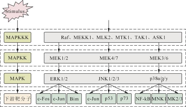
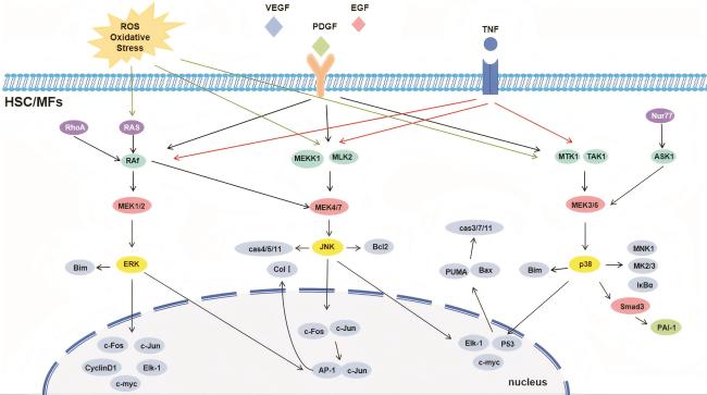
 Fig 1 Schematic representation of the MAPK signaling cascadeFig 2 Schematic representation of the MAPK signal transduction network and its molecular regulatory mechanisms
/
| 〈 |
|
〉 |

